Primer Dimer Checker for quick PCR review
A Primer Dimer Checker helps you find primer-primer binding risk before PCR, qPCR, cloning PCR, or sequencing PCR. It compares the forward primer and reverse primer for simple complementary regions. It pays special attention to the 3′ end because DNA polymerase extends from that end.
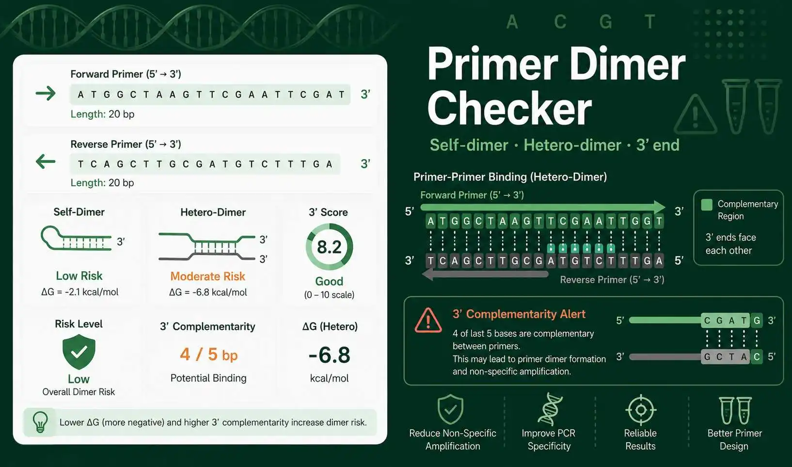
Primer dimers form when primers bind to each other instead of binding only to the target DNA template. The reaction can then amplify a small unwanted product. This can reduce yield, create extra gel bands, distort qPCR fluorescence, and make melt curves harder to interpret.
What this primer dimer tool checks
The tool accepts two DNA primers written in the 5′ to 3′ direction. It removes spaces, line breaks, numbers, and FASTA headers. It then checks forward self-dimer, reverse self-dimer, and forward-reverse hetero-dimer signals.
Each result includes the longest contiguous complementary run, a 3′ complementarity score, total matching bases in the strongest simple alignment, and a low, moderate, or high risk label. The same screen also shows primer length, GC percentage, 3′ GC clamp, estimated Tm, average Tm, and Tm difference.
Primer dimer score meaning
A short complementary region inside a primer is common. It does not always cause a failed reaction. A longer complementary run is more concerning, especially when it includes the 3′ terminal bases. A 3′ match can act like a tiny primed template and allow polymerase extension.
The risk label is a practical screen, not a thermodynamic prediction. A high score means you should redesign or verify the primer pair. A moderate score means you should inspect the alignment and your assay conditions. A low score means this simple sequence check did not find a strong dimer signal.
Primer dimer equations and simple rules
This checker uses sequence complementarity. A pairs with T. C pairs with G. The tool creates the reverse complement of a primer and scans for matching regions against the other primer sequence.
GC content is calculated with this equation: GC% = (G + C) ÷ total bases × 100. Tm is estimated with a basic educational primer formula. For short primers, the Wallace rule is used: Tm = 2(A + T) + 4(G + C). For longer primers, the tool uses a simple length-adjusted estimate.
Thermo Fisher primer design guidance recommends avoiding primer self-complementarity and forward-reverse complementarity because those patterns can lead to hairpins, self-dimers, or primer-dimers.Thermo Fisher PCR primer design tips
Worked example for primer-dimer checking
Suppose the forward primer is ATGCGTACGTTAGCGTACGA and the reverse primer is TCGTACGCTAACGTACGCAT. The checker first cleans the sequences. It then calculates length, GC content, Tm, and 3′ GC bases.
Next, it compares each primer with its own reverse complement to look for self-dimer risk. It also compares the forward primer with the reverse complement of the reverse primer to look for hetero-dimer risk. If the strongest warning is at the 3′ end, the result becomes more important than an internal match of the same size.
In a lab notebook or student report, you can write: “The primer pair was screened for self-dimer and hetero-dimer formation. The main values reviewed were GC content, Tm difference, longest complementary run, and 3′ complementarity score.”
Use case 1: checking PCR primers before ordering
Use this tool after you choose primers for a PCR amplicon. Paste both primer sequences and review the dimer risk label. If the forward + reverse dimer result is high, redesign one primer. Pay close attention when both primers have complementary 3′ ends because that pattern can produce primer-dimer bands.
Also compare the Tm values. A large Tm difference can make one primer bind more efficiently than the other. If you need a temperature-focused check, compare this output with the Primer Tm Calculator.
Use case 2: reviewing qPCR primer pairs
qPCR assays are sensitive to primer-dimers because small unwanted products can still bind fluorescent dyes. A primer-dimer product may appear as a late Ct signal or an extra melt curve peak. Use this checker before running qPCR, especially when a no-template control gives amplification.
This tool does not judge qPCR efficiency by itself. After dimer screening, use a standard curve, melt curve, and no-template control to confirm assay behavior. You can also use the Oligo Analyzerfor a broader primer property check.
Practical problem: decide whether to redesign
Imagine a primer pair has 48% GC content, a 2°C Tm difference, and a high 3′ complementarity score. The GC and Tm values look acceptable, but the 3′ score still matters. The best decision is to redesign one primer or move the primer location slightly on the template.
Now imagine another pair has a moderate internal complementarity score but no strong 3′ match. That pair may still work, especially if the assay has clean specificity. You should verify it with a more detailed tool and real PCR controls instead of rejecting it automatically.
Common mistakes when checking primer dimers
Do not paste the reverse complement of the reverse primer unless your protocol specifically asks for it. Most primer sequences should be entered exactly as ordered, from 5′ to 3′. The tool handles reverse complement comparisons internally.
Do not judge primers by one value only. Primer design also depends on target specificity, amplicon length, template GC richness, annealing temperature, polymerase, magnesium concentration, and reaction conditions.
What to verify before real PCR use
Verify primer specificity with the target sequence, confirm expected product size, check both primer Tm values, inspect the 3′ ends, and run proper controls. For important experiments, confirm dimer and hairpin predictions with a validated thermodynamic tool before placing an oligo order.